- The Laboratory
- Organization
- Departments
- Jobs
- Analysis book
- Contact
- News
- Publications
- Download


The Breast Cancer Unit of the CHL Kriibszentrum, which has valued the quality of interdisciplinary oncological care facilities for over 10 years, in partnership with l’Institut National du Cancer (INC) has this year been awarded the OnkoZert certificate of the German Cancer Society (DKG). This internationally recognised certification is thus an appreciation of the excellent treatment options for patients in Luxembourg and certifies holistic and comprehensive care from the time of diagnosis to long-term aftercare. The CHL’s breast cancer department works closely with the Laboratoire national de santé and the Centre François Baclesse. Without the cooperation of these three institutions, such high-level certification would not have been possible.
The Laboratoire national de santé (LNS) with its two national diagnostic centres, the National Centre of Pathology (NCP) and the National Centre of Genetics (NCG), is a principal actor in the field of cancer diagnosis and screening. The NCP’s pathology service deals with the diagnosis of cancers and pre-cancerous lesions, inflammatory lesions as well as pseudo-tumoral lesions and malformations in the various organs of a patient. It also relies on molecular diagnostics of tumours, in collaboration with the NCG’s Molecular Genetics Unit. The NCG has been offering comprehensive medical care for hereditary disorders since 2018, including clinical genetic diagnostics, genetic counselling, and genetic testing to the Luxembourgish population. This also includes genetic diagnosis and counselling for patients and families in the context of inherited cancer predisposition syndromes, such as Hereditary Breast and Ovarian Cancer Syndrome (HBOC).
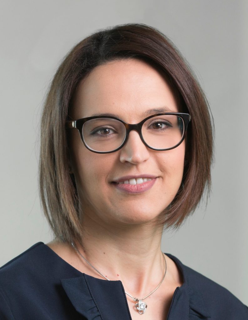
Dr Olfa Chouchane Mlik has headed the Service of Anatomical Pathology at the NCP since January 2018. Her team was instrumental in CHL seeing itself awarded the OnkoZert accreditation, thanks to the work of the team’s tumour boards. These interdisciplinary expert panels meet regularly to discuss the best possible treatment for patients. Below, Dr Mlik provides us with an insight into her unit and its role in breast cancer screening and treatment.
| Anatomopathology (or anatomical pathology) is a specialty that is little-known by the general public. Cell and tissue samples taken during a radiological, endoscopic, or surgical examination are scrutinised by pathologists under a microscope. Cancer is a disease characterised by the uncontrolled development and proliferation of abnormal cells that have escaped all the body’s control and repair mechanisms. The role of the pathologist is to detect the tissue deformation caused by this disease, to recognise its type, form, and degree of aggressiveness and to characterise it, if necessary, by means of biomarkers which also have a prognostic value. This information, which the pathologist will have passed on to all the doctors concerned, will then determine the treatment offered to the patient. The “anapath” report is a checklist to help the surgeon, the oncologist and the radiotherapist who will intervene to complete the treatment. |
 Dr Olfa Chouchane Mlik |
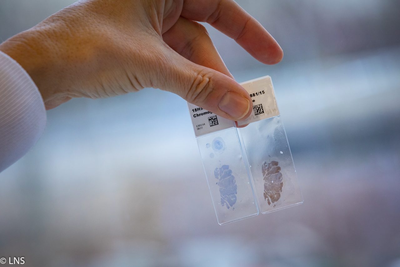
In the LNS’ anatomical pathology unit, we process an average of 250 samples per day. Tens of thousands of thin sections (of the order of one micron) on glass slides are thus taken, with tissue or cytological samples frozen in formalin and packaged in paraffin, in order to preserve them for decades. These sections are then stained and put under the microscope by the department’s pathologists. The pathologist may also be called upon during surgery to carry out a rapid analysis (extemporaneous examination) in order to clarify a clinically suspicious diagnosis or to confirm with the surgeon that a tumour has been completely removed. In our profession, it is important to remember that behind every histological section examined, a person is anxiously waiting for the result and the diagnosis. Therefore, in case of doubt, our teams often gather together around the microscope to discuss histological section analyses that aren’t clear cut.
Our pathologists are also available to provide patients with more detailed information on their diagnosis and explain open questions in a face-to-face meeting. The contact details of the treating pathologist are indicated on the corresponding medical report.
Advances in research have led to a better understanding of the biological mechanisms behind the development and progression of cancers. We now know that is not a case of cancer per organ but several subtypes of cancer, each with different abnormalities. We know that the more precise the level of information provided in the post-examination report, the more we will be able to adjust the treatments offered to patients in a targeted and personalised manner. Precision medicine starts with the pathological analysis of the tumour and is enriched by molecular analysis, which makes it possible to identify other biological anomalies that can be targeted by specific treatments.
This is why, for example, the tumour tissue is tested for mutations in genes involved in DNA repair, in particular BRCA genes. This process requires the expertise and cooperation of the National Centre of Pathology and the National Centre of Genetics. Patients with a mutation in one of the BRCA genes may be eligible to treatment with PARP inhibitors. PARP inhibitors (iPARP) are anti-cancer molecules belonging to the targeted therapy family. They can be offered in the treatment of metastatic breast cancer linked to a BRCA mutation. Furthermore, if a mutation is identified in the tumour, the patient and families are referred for genetic counselling and further genetic testing to the NCG to confirm if a hereditary cancer predisposition syndrome is present in the family.
Each year between 400 and 450 new cases of breast cancer are diagnosed in Luxembourg. The national breast cancer screening programme “mammography programme” has existed since 1992. It is aimed at all women aged between 50 and 70. A mammogram is carried out every two years and allows breast cancer to be detected at an early stage (before any clinical signs). Before the age of 40, only women at high risk of cancer are invited for individual follow-up with a specialised imaging assessment.
I also encourage women to self-check as nobody knows our body better than ourselves. While not a substitute for clinical examination or mammography screening, breast self-examination can allow us to detect changes that could lead to a diagnosis of cancer. Obviously, any suspicion raised during a breast self-examination warrants a specialist opinion without delay.
We work with all the players in the Luxembourg health sector (hospitals, clinics, and private practices) on a daily basis and we actively participate in the various projects for the implementation of cancer management guidelines coordinated by the National Cancer Institute, such as the “national guidelines for the diagnosis and treatment of gynaecological cancers” and the “guidelines for patient care for breast cancer”.
We also collaborate with other pathology services in cities in neighbouring countries (Liege, Brussels, Cologne, Paris), either in the context of external quality control or for additional expertise recommended in certain rare pathologies. We are also actively involved in research projects with partners both in Luxembourg (Luxembourg Institute of Health, etc.) and internationally (Institut Curie in Paris, Institut national de la santé et de la recherche médicale – INSERM, etc.). Our vision is to deliver a service of excellence to all patients in the Luxembourg region, in line with international standards, as part of a continuous improvement process.
A webinar with Dr Olfa Chouchane Mlik on the role of the pathologist in management of breast biopsy is available on the “Groupe Sein” Blog of the Centre Hospitalier du Luxembourg (CHL) recorded as part of Pink October: https://www.chl.lu/fr/actualites/octobre-rose-au-chl-2021-table-ronde-1. The blog plays a key role in providing useful information and offers a platform for patients and their families to share their experiences.

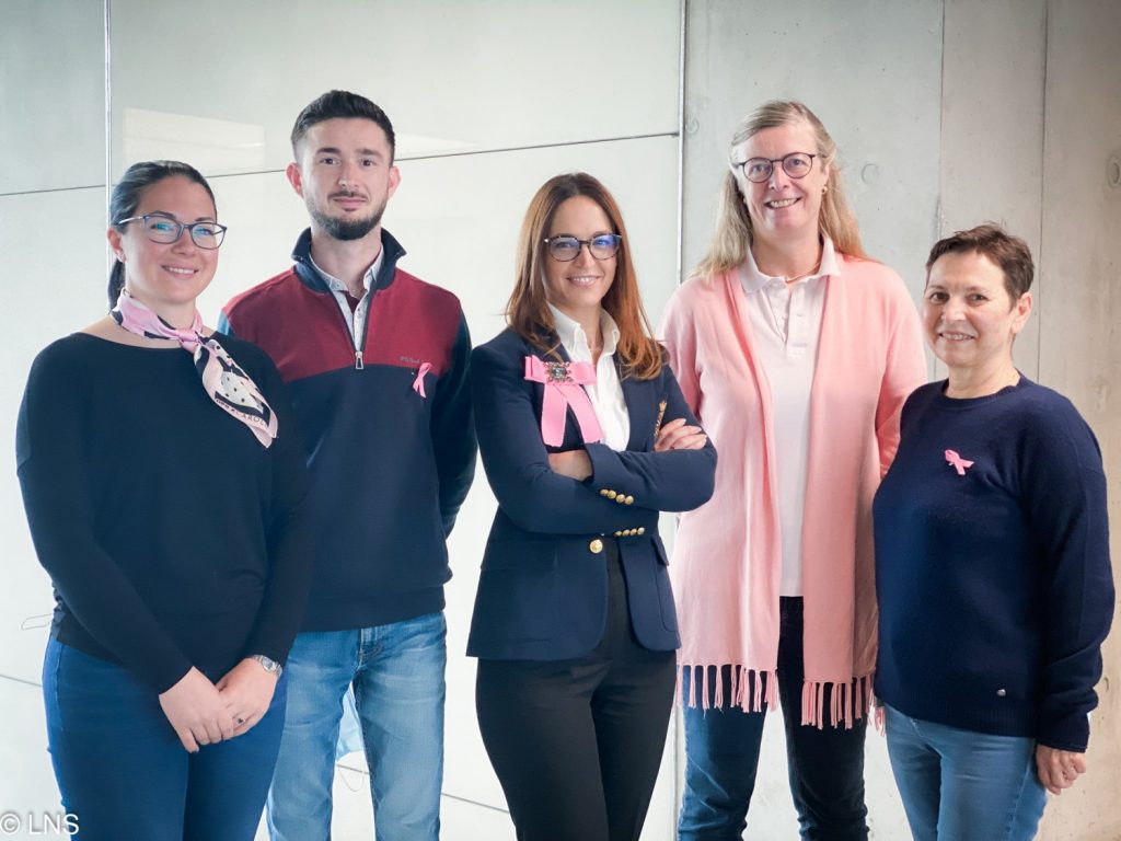
Dr Olfa Chouchane Mlik entourée de son équipe du NCP